Cardiac Imaging
May 2023
EN.510.433 Medical Image Analysis
Objectives:
Skills Applied:
Comments:
The project was largely open-ended, allowing me to apply any image analysis technique from course materials. This project adopted classic computer vision techniques , rather than ML-based models, where I gained a much deeper understanding of their practical applications. In the future, I'm curious about ML-based approachesas well, where the model setting accounts for a-priori constraints.
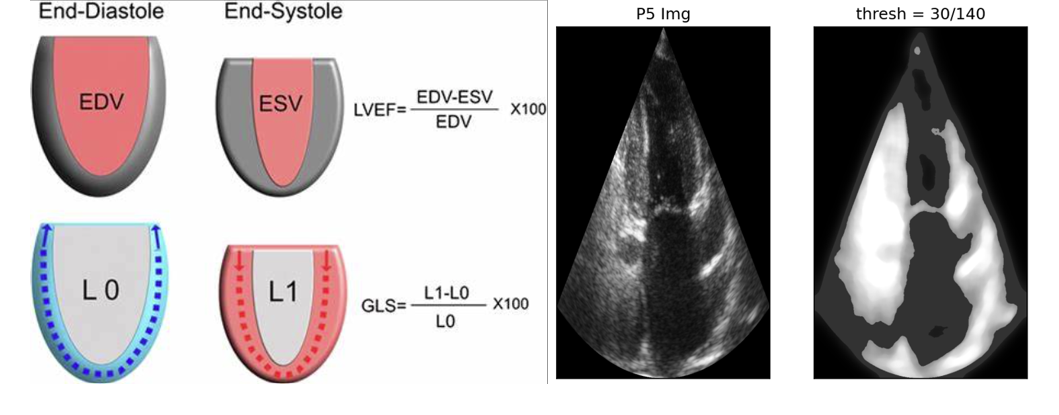
Left: formulas for end-diastolic Volume (EDV), end-systolic volume (ESV), and ejection fraction (EF). Right: image pre-processing
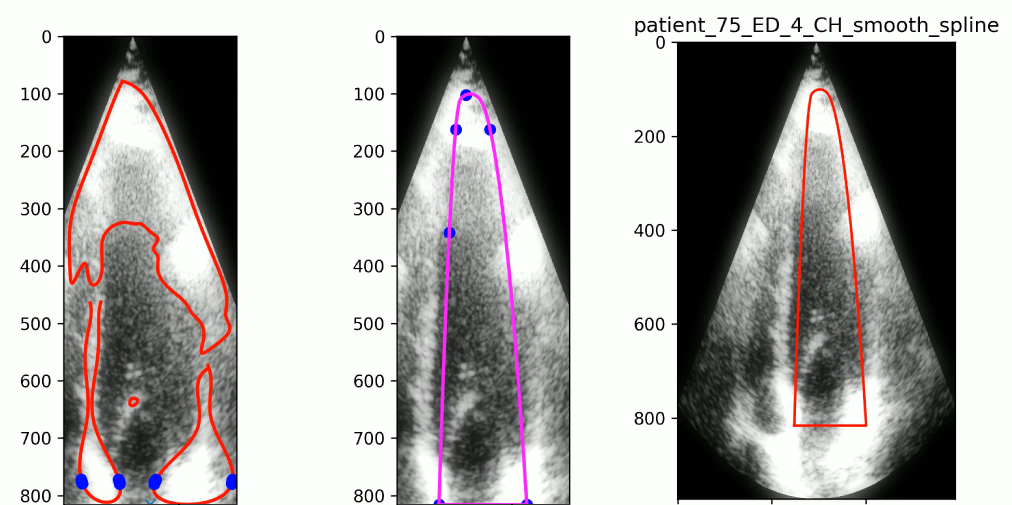
Region of interest labeling
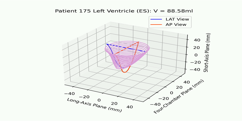
ESV reconstructed video
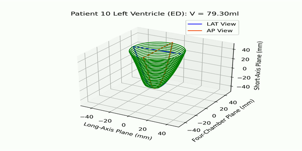
EDV reconstructed video